Beranda
/ Shoulder Ligament Anatomy Diagram / Anterior Aspect Of The Shoulder Including Ligaments And Bursa Shoulder Anatomy Joints Anatomy Shoulder Joint Anatomy : 7 draw labelled diagram showing the relations of shoulder joint.
Shoulder Ligament Anatomy Diagram / Anterior Aspect Of The Shoulder Including Ligaments And Bursa Shoulder Anatomy Joints Anatomy Shoulder Joint Anatomy : 7 draw labelled diagram showing the relations of shoulder joint.
Insurance Gas/Electricity Loans Mortgage Attorney Lawyer Donate Conference Call Degree Credit Treatment Software Classes Recovery Trading Rehab Hosting Transfer Cord Blood Claim compensation mesothelioma mesothelioma attorney Houston car accident lawyer moreno valley can you sue a doctor for wrong diagnosis doctorate in security top online doctoral programs in business educational leadership doctoral programs online car accident doctor atlanta car accident doctor atlanta accident attorney rancho Cucamonga truck accident attorney san Antonio ONLINE BUSINESS DEGREE PROGRAMS ACCREDITED online accredited psychology degree masters degree in human resources online public administration masters degree online bitcoin merchant account bitcoin merchant services compare car insurance auto insurance troy mi seo explanation digital marketing degree floridaseo company fitness showrooms stamfordct how to work more efficiently seowordpress tips meaning of seo what is an seo what does an seo do what seo stands for best seotips google seo advice seo steps, The secure cloud-based platform for smart service delivery. Safelink is used by legal, professional and financial services to protect sensitive information, accelerate business processes and increase productivity. Use Safelink to collaborate securely with clients, colleagues and external parties. Safelink has a menu of workspace types with advanced features for dispute resolution, running deals and customised client portal creation. All data is encrypted (at rest and in transit and you retain your own encryption keys. Our titan security framework ensures your data is secure and you even have the option to choose your own data location from Channel Islands, London (UK), Dublin (EU), Australia.
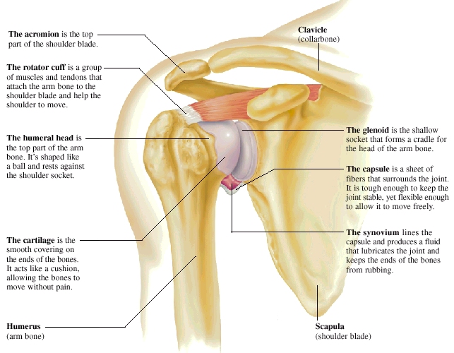
Shoulder Ligament Anatomy Diagram / Anterior Aspect Of The Shoulder Including Ligaments And Bursa Shoulder Anatomy Joints Anatomy Shoulder Joint Anatomy : 7 draw labelled diagram showing the relations of shoulder joint.. Corey chakarun from shin imaging in california. Radiologists primarily perform shoulder imaging to assess injuries within the shoulder joint. Understanding shoulder anatomy can help to avoid injury, promote rehabilitation, and can assist you in using the joint optimally. The shoulder is one of the largest and most complex joints in the body. It is the major joint connecting the upper limb to the trunk.
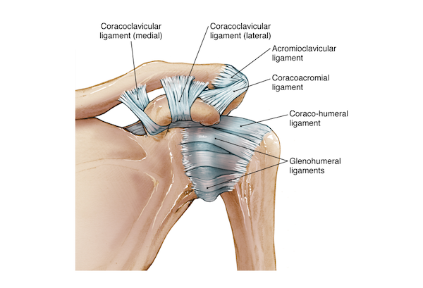
You can see it enclosing the glenohumeral joint and you can see its attachment on the anatomical you've got the transverse humeral ligament and the coracohumeral ligament. 7 draw labelled diagram showing the relations of shoulder joint. Divided into two additional ligaments including the trapezoid ligament. Radiologists primarily perform shoulder imaging to assess injuries within the shoulder joint. An image depicting shoulder anatomy can be seen below.
Shoulder Anatomy from ix-cdn.b2e5.com You can see it enclosing the glenohumeral joint and you can see its attachment on the anatomical you've got the transverse humeral ligament and the coracohumeral ligament. Robin smithuis and henk jan van der woude. The shoulder joint is formed where the humerus (upper arm bone) fits into the scapula. Shoulder joint of human body anatomy infographic diagram with all parts including bones ligaments muscles bursa cavity capsule cartilage membrane for medical science education and health care. There are several important ligaments in the shoulder. The shoulder joint (glenohumeral joint) is a ball and socket joint between the scapula and the humerus. One or more ligaments provide stability to a joint during rest and movement. This page is about shoulder anatomy ligaments and muscles,contains soft tissues of the shoulder,shoulder joint;
It is the major joint connecting the upper limb to the trunk.
There are five major shoulder ligaments that keep the shoulder in place and prevent it from dislocating. There are several important ligaments in the shoulder. The transverse humeral ligament is not shown on this diagram. Shoulder anatomy diagram / normal shoulder anatomy. Start studying shoulder ligaments and tendons. Additional stability is provided by: We hope you will use this picture in the study and helping contents of axilla diagram. The shoulder joint is formed where the humerus (upper arm bone) fits into the scapula. Understanding shoulder anatomy can help to avoid injury, promote rehabilitation, and can assist you in using the joint optimally. Rotator cuff & scapula stabilising. The human shoulder is made up of three bones: Simply put, the shoulder, or shoulder joint, is the connection of the upper arm and the thorax. Webmd's shoulder anatomy page provides an image of the parts of the shoulder and describes its function, shoulder problems, and more.
Static:gh ligaments, labrum & capsule and dynamic constraints: This mr arthrogram of the shoulder was performed on a normal male patient on a ge signa pioneer 3t mri by dr. Robin smithuis and henk jan van der woude. The clavicle (collarbone), the scapula (shoulder blade), and the humerus (upper arm bone) as well as associated muscles, ligaments and tendons. Shoulder and back muscles diagram.
Common Injuries Of The Shoulder from www.stanfordchildrens.org One or more ligaments provide stability to a joint during rest and movement. Although the joint is held together by these extensive ligament and muscle attachments, certain types of forces can weaken the shoulder easily. The primary function of the shoulder girdle is to give strength and range of motion to the arm. There are several important ligaments in the shoulder. (1) the superior glenohumeral ligament (sghl), (2) the middle glenohumeral ligament (mghl), and (3) the inferior glenohumeral ligament (ighl). Ligaments are fibrous bands or sheets of connective tissue linking two or more bones, cartilages, or structures together. Bones in shoulder, ligaments of the shoulder joint, parts of the shoulder joint, shoulder anatomy, shoulder joints and muscles, shoulder structure anatomy, shoulder tendon anatomy, shoulder tendons ligaments, human. Simply put, the shoulder, or shoulder joint, is the connection of the upper arm and the thorax.
The shoulder is one of the largest and most complex joints in the body.
One or more ligaments provide stability to a joint during rest and movement. Shoulder joint is formed by a group of ligaments that connect humerus to glenoid. Additional stability is provided by: The disk has a great variation in size and shape. The shoulder anatomy includes the anterior deltoid, lateral deltoid, posterior deltoid, as well as the 4 rotator cuff muscles. Glenohumeral joint,shoulder tendons,8 ejercicios para el hombro que debemos hacer and more. It can help you understand our world more detailed and specific. Simply put, the shoulder, or shoulder joint, is the connection of the upper arm and the thorax. Start studying shoulder ligaments and tendons. The most common shoulder injuries involve the muscles, ligaments, cartilage, and tendons, rather than the bones. Webmd's shoulder anatomy page provides an image of the parts of the shoulder and describes its function, shoulder problems, and more. Home > blog > anatomy > shoulder anatomy: The glenohumeral ligaments can be seen here, but they're not really.
Shoulder anatomy diagram / normal shoulder anatomy. Learn about shoulder anatomy from darren keiser. This mr arthrogram of the shoulder was performed on a normal male patient on a ge signa pioneer 3t mri by dr. The transverse humeral ligament is not shown on this diagram. Superior glenohumeral ligament and coracohumeral ligament are the primary restraints to posterior translation with the are flexed, adducted and internally acromioclavicular ligament anatomy.
Shoulder Anatomy from uploads-ssl.webflow.com The shoulder is not a single joint, but a complex arrangement of bones, ligaments, muscles, and tendons that is better called the shoulder girdle. A joint capsule is a watertight sac that surrounds a joint. We hope you will use this picture in the study and helping contents of axilla diagram. The transverse humeral ligament is not shown on this diagram. Shoulder and back muscles diagram. Robin smithuis and henk jan van der woude. The human shoulder is made up of three bones: Notice superior labrum and attachment of the superior glenohumeral ligament.
Notice superior labrum and attachment of the superior glenohumeral ligament.
Last update february 25, 2021. The glenohumeral ligaments can be seen here, but they're not really. You can see it enclosing the glenohumeral joint and you can see its attachment on the anatomical you've got the transverse humeral ligament and the coracohumeral ligament. Rotator cuff & scapula stabilising. The disk has a great variation in size and shape. This diagram here just shows the joint capsule itself. Robin smithuis and henk jan van der woude. Coronal section of shoulder joint. The shoulder is not a single joint, but a complex arrangement of bones, ligaments, muscles, and tendons that is better called the shoulder girdle. Anatomy is the amazing science. Superior glenohumeral ligament and coracohumeral ligament are the primary restraints to posterior translation with the are flexed, adducted and internally acromioclavicular ligament anatomy. We hope you will use this picture in the study and helping contents of axilla diagram. Learn vocabulary, terms and more with flashcards, games and other study tools.
The disk has a great variation in size and shape shoulder anatomy diagram. These ligaments are main source of stability for the shoulder.